Surface EMG Technology
What are the specific principles and procedures of surface diaphragmatic EMG?
Surface diaphragmatic electromyography (sEMGdi) is a non-invasive technique used to monitor the electrical activity of the diaphragm, the primary muscle involved in breathing. Electrodes placed on the skin over the lower rib cage detect signals from the diaphragm where it attaches to the ribs.
Muscle signals are inherently noisy signals because they are the sum of individual muscle activation spikes, which when grouped together and shown as the root mean square show the overall activation of the muscle over time.
By using sEMGdi to monitor the patient over time, clinicians can analyze trends such as:
- Slope of the inspiration curve
- Amplitude levels
- Area under the curve
Additionally using end-expiratory occlusion maneuvers at various levels of positive end-expiratory pressure (PEEP) the neuromechanical coupling index can be calculated (as the ratio of end-expiratory occlusion pressure (Pocc) to sEAdi). This index helps assess changes in the patient’s cardiorespiratory condition, for example if the patient is getting weaker, stronger or responding to certain treatments.
Where should electrodes be placed to measure diaphragmatic EMG activity?
For neonatal patients: please see the clinical article from R.W. van Leuteren, R.E. Bekhuis, C.G. De Waal, F.H. de Jongh, A.H. van Kaam, G.J. Hutten, Diaphragmatic electromyography in preterm infants: The influence of electrode positioning, Pediatric Pulmonology. 2019;1–6. DOI: 10.1002/ppul.24585.
For adults;
- (Black) reference electrode: Somewhere on the center of the chest (sternum).
- (White) measurement electrodes (2x): on the diaphragm (lower rib cage) on the right and left side of the body.
How to find the diaphragm?
Start from the nipple and move vertically down to the intersection of the last rib and abdominal muscles. Place the electrode half on the rib and half on the abdomen; slightly higher is better than too low. This ensures optimal detection of diaphragmatic electrical signals.
How do you know that the electrodes are positioned on the right place?
Correct electrode placement is essential for capturing accurate surface electromyography (sEMG) signals from the muscles of interest.
- For intercostal muscles (between the ribs):
Electrodes are placed directly on the skin above the target muscle. - For diaphragmatic muscles (located deeper and behind the rib cage):
Electrodes should be positioned near where the diaphragm attaches to the lower ribs. Although the diaphragm lies deeper, its electrical activity can still be detected through the skin when electrodes follow the curve of the lower rib cage.
Electrode placement is checked by ensuring it aligns with the physiological contour of the lower ribs. This anatomical guidance helps target the diaphragm effectively.
Even with correct placement, the quality of the sEMG signal depends on:
- The patient’s physiological muscle activity.
- The EMG device’s filter settings.
What is the normal range for surface diaphragmatic EMG signal amplitude?
The amplitude of surface diaphragmatic EMG (sEMG) signals can vary significantly between individuals due to physiological differences such as muscle development, genetic predisposition, and underlying health conditions.
- Typical baseline (tonic) activity between breaths: Approximately 5 µV.
- Peak amplitudes during active breathing: Can reach 25 µV or higher, depending on the individual’s effort and muscle activation.
If clear breathing patterns are not visible in the sEMG signal, consider checking the following:
- Electrode placement: Ensure electrodes are positioned correctly along the lower rib cage, following the physiological curve.
- Patient posture: The patient should be lying down, relaxed, and not speaking.
- Abdominal movement: Minimize motion artifacts by ensuring the abdomen remains still during measurement.
What techniques can be used to validate the correlation between surface diaphragmatic EMG signals and diaphragmatic activity?
To validate the correlation between surface diaphragmatic EMG (sEMG) signals and actual diaphragmatic activity, we currently apply two complementary techniques:
- Ultrasound Imaging
We have recently begun investigating how diaphragmatic EMG signals relate to diaphragmatic movement and thickness using ultrasound. This allows us to visually assess diaphragm dynamics and compare them with the electrical activity recorded by sEMG. - Flow and Pressure Measurements
In ongoing studies using the SERA device, we incorporate a digital flow/pressure sensor into the measurement setup. This enables synchronous data collection from both the SERA device and the flow/pressure sensor. The combined data is output as a single package to the PC, allowing for direct comparison between EMG signals and respiratory parameters such as airflow and pressure.
These methods help assess whether the recorded EMG signals accurately reflect diaphragmatic function.
Are there any notable differences in sEMG signals across age groups, genders, or ethnicities?
So far, we have not observed any notable differences in surface EMG (sEMG) signals between different age groups or ethnicities. However, we do see significant individual variation, which is often related to developmental differences and the clinical situation of the patient.
In general:
- Spontaneously breathing neonates tend to show higher sEMG signal amplitudes, as they need to exert more effort to open their lungs.
- Healthy adults, on the other hand, typically show lower sEMG signals, since their breathing requires less muscular effort.
It’s important to note that even among healthy adults, sEMG signals can vary due to differences in how their respiratory muscles have developed over time.
Could sEMG data be used to retroactively assess whether diaphragmatic motion is active or passive?
In the ongoing clinical trial using the Macawi SERA device, the work of breathing is measured at varying levels of non-invasive positive pressure ventilation in neonates. Initial, non-conclusive results show that increased respiratory support leads to a decrease of the patients work of breathing, indicating less activity by the diaphragm muscle, suggesting a shift toward passive motion.
In contrast, other clinical trials involving spontaneously breathing patients have shown a clear increase in diaphragmatic activity and Work of Breathing during events such as obstructive apnea.
These findings suggest that it is likely possible to distinguish between active and passive diaphragmatic motion based on Work of Breathing information derived from sEMG measurements.
What is the accuracy of non-invasive diaphragmatic EMG (sEMG)?
The accuracy of surface diaphragmatic EMG (sEMG) remains a key focus for our team. We are about to begin a study investigating the correlation between ultrasound measurements of the diaphragm and sEMG signals. Our long-term goal is to develop a wearable ultrasound patch in combination with sEMG to help optimize ventilator settings for patients in intensive care. We look forward to sharing our findings once preliminary results become available.
In the meantime, we can provide sample data showing the correlation between sEMG and flow/pressure measurements. Please let us know if you would like to receive this information.
We would also be very interested in hearing about your findings.
Macawi SERA Functionality and specifications
What type of signal does the macawi SERA sensor use?
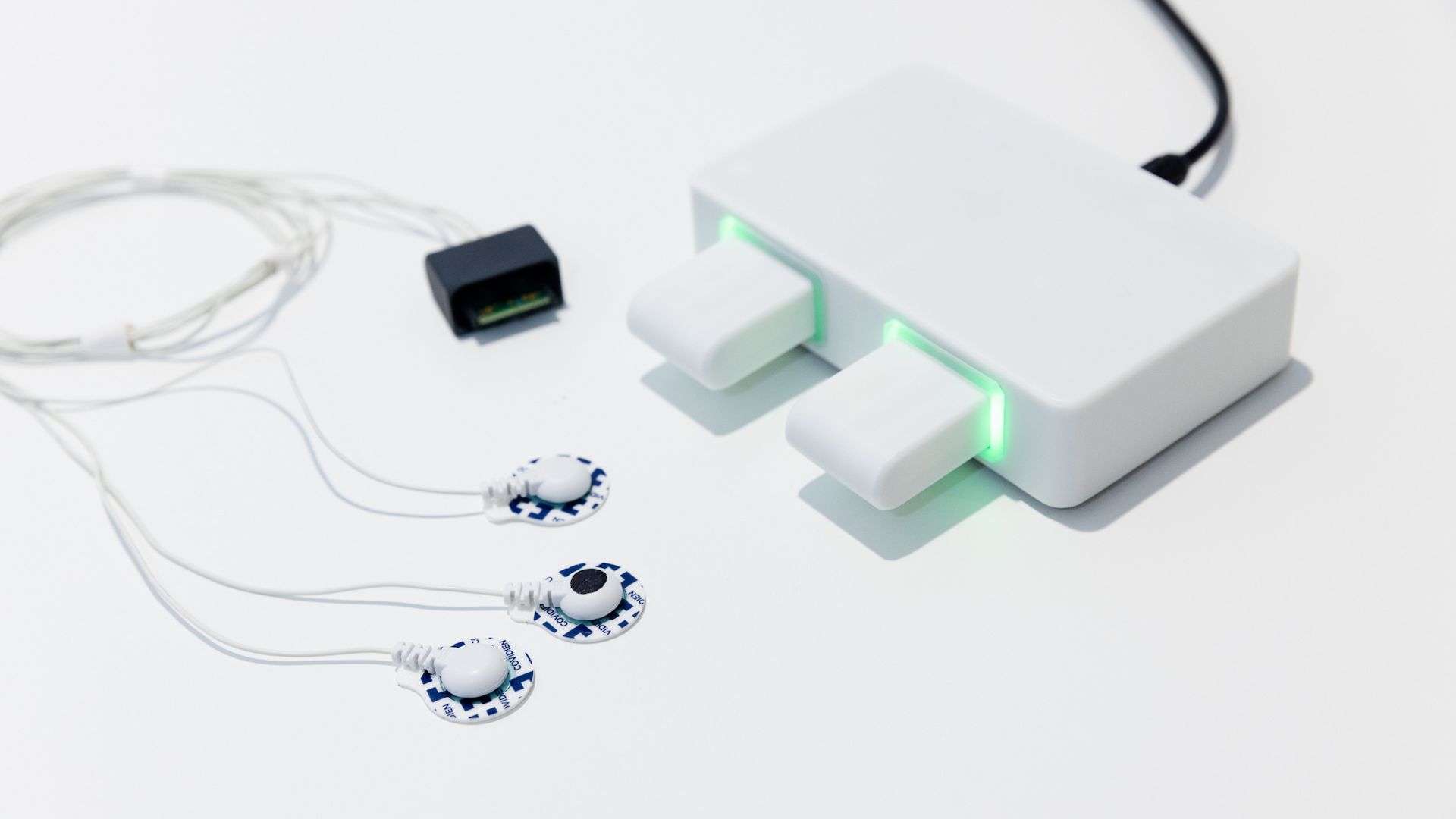
The Macawi SERA sensor measures a transcutaneous electromyographic signal of the diaphragm, commonly referred to as dEMG. The system uses hydrogel electrodes placed on the skin above the muscle group of interest. These electrodes detect the electrical activity generated by the activation potentials (spikes) of individual muscle fibers. Since this raw signal is inherently noisy, the SERA device calculates the root mean square (RMS) of the electrical potential. This RMS value represents the average electrical activity of the targeted muscle group over time, providing a stable and interpretable signal for monitoring diaphragmatic function.
How sensitive is the Macawi SERA system to EMG signals?
The Macawi SERA system is highly sensitive to muscle activity, with a baseline noise level of approximately 1 µV RMS. This means that any muscle signals stronger than 1 µV RMS can be clearly detected, while signals below this threshold may be lost in the noise. To interpret the inherently noisy EMG signal, the system calculates the root mean square (RMS) of the electrical potential, providing a reliable measure of overall muscle activity.
Is skin preparation required or different across patient groups?
- Hydrogel electrodes are used with the Macawi SERA and do not require skin preparation. The hydrogel ensures good contact with the skin across all patient groups: from premature infants to adults.
- If other types of electrodes (e.g., dry electrodes) are used, skin preparation such as cleaning with alcohol wipes or gentle abrasion may be necessary. However, we have limited experience with these alternatives and recommend using hydrogel electrodes for optimal performance.
Is there a left/right distinction when using two electrodes and one reference with the Macawi SERA system?
No, there is no left/right distinction between the two white electrodes used with the Macawi SERA system. Both electrodes are identical, and switching their positions at the start of a measurement does not affect the signal quality or accuracy. The system is designed to measure the differential electrical activity from the muscle group beneath the electrodes, which is obtained by subtracting the signal of one of the electrodes from the other, thereby making it irrelevant which side each electrode is placed on.
How does the Macawi SERA distinguish between abdominal and diaphragmatic muscle activity?
The SERA system cannot directly differentiate between electrical activity from the diaphragm and abdominal muscles. All muscle groups produce similar EMG signal profiles, making it difficult to isolate one from the other based solely on signal characteristics.
To improve specificity:
- Electrode placement is key. Positioning electrodes closer to the rib cage and away from the abdomen helps reduce signal contamination from abdominal muscles.
- Patient selection and behavior also matter. Patients who are actively using their abdominal muscles, due to movement, talking, or other activity, are not ideal candidates for accurate sEMG measurement.
This challenge is not unique to sEMG. Other respiratory monitoring technologies, such as impedance-based sensors and mechanical movement sensors (e.g., Graseby belts or Respibands), also experience interference from non-respiratory muscle activity.
How sensitive is Macawi SERA compared to the Graseby capsule in monitoring respiratory events?
Macawi SERA and the Graseby capsule measure different aspects of the breathing process:
- Macawi SERA detects the electrical activity of the diaphragm, capturing the muscle activation that initiates breathing.
- Graseby capsule measures the mechanical expansion of the chest, which is the result of diaphragmatic contraction.
In this sense, SERA measures the actant, while Graseby measures the resultant. Because muscle activation precedes chest expansion, the SERA signal typically appears slightly earlier than the Graseby signal. Despite measuring different physical properties, the two signals are often highly correlated—stronger diaphragmatic contractions lead to greater chest expansion.
However, in cases of airway obstruction, this correlation breaks down:
- The diaphragm may still contract (visible in the EMG signal),
- but air cannot enter the lungs, so the chest does not expand. Resulting in no signal from the Graseby capsule.
Sensitivity Considerations
The sensitivity of both systems is patient-dependent:
- Patients with stiff lungs or chest walls may show high EMG amplitudes (due to strong muscle effort) but low Graseby signals (limited chest expansion).
- Patients with compliant lungs and well-developed muscles may show lower EMG amplitudes and higher Graseby signals.
Are the raw and unfiltered EMG and ECG signals also recorded, as well as the filtered/processed signals?
Yes, Macawi SERA provides raw unfiltered as well as filtered signal information.
Can the signals from Macawi SERA be downloaded to a CSV?
Yes, when using the Macawi SERA system with a computer and the provided R&D graphical user interface (GUI), it is straightforward to log incoming data streams in CSV format. The CSV file includes:
- Raw ExG data from the electrodes
- Processed ECG and EMG waveforms
- Breath-by-breath analysis parameters
For data logging when the SERA system is connected to a hospital monitor, please refer to the monitor’s user manual or consult the hospital’s ICT department, as integration and data extraction methods may vary by system.
How many muscle groups can be measured simultaneously using Macawi SERA?
The number of muscle groups that can be measured simultaneously with the Macawi SERA system depends on the number of electrode leads available. Typically, a muscle group is characterized using the differential signal between two leads.
- A 3-electrode system can measure 1 muscle group
- A 5-electrode system can measure 2 muscle groups
- A 7-electrode system can measure 3 muscle groups
Each additional pair of electrodes allows for the monitoring of an additional muscle group, assuming proper placement and signal quality.
Can generic ECG electrodes be used instead, when the originally provided ones are running out?
Yes, you can use any commercially available ECG snap electrode with the Macawi SERA system. These electrodes are compatible with the SERA snappers and can serve as an alternative when the supplied electrode patches are running low.
For optimal signal quality, we recommend continuing to use hydrogel-based electrodes, as they provide reliable skin contact without requiring additional preparation.
Is it possible to integrate the Macawi SERA sensor into a ventilator? If so, how does it communicate, and can the communication protocol and electrical diagrams be accessed?
Yes, the Macawi SERA sensor can be integrated into a ventilator system.
- Communication:
The SERA device communicates via a digital signal using a defined communication protocol. This protocol outlines how data can be received from the device. - Integration Requirements:
While the electrical diagram of the SERA system is not publicly available, we provide an integration guideline document. This document specifies the necessary hardware on the host side, including:- A compatible connector
- An RS232 transceiver
- An electrical isolation barrier
- Technical Specifications:
These include the electrical properties of the digital communication signal and are available to support integration efforts.
If you're planning integration, we recommend reaching out to request the integration guideline and technical specifications directly.
Can Macawi SERA be connected to any hospital monitor, and are special software or drivers required?
This is possible in close collaboration with manufacturers of patient monitoring equipment.
The Macawi SERA outputs digital serial data at 1 Mbaud using RS232 signal levels and a defined communication protocol. To connect SERA to a patient monitor, the following hardware must be present on the monitor side:
- A compatible connector
- An RS232 transceiver
- An electrical isolation barrier
In addition, the monitor must be able to communicate with the SERA device using its protocol to receive and display data. No special drivers are required from Macawi, but custom software or firmware on the monitor may be needed to interpret and visualize the incoming data correctly.
What are some of the technical specifications of the Macawi SERA? (for example the signal-to-noise ratio, the resolution, how well it removes ECG artefacts, etc)
For full details, please refer to the specification sheet available on our webpage.
Core technical specifications
- Sample rate: 500 Hz
- Input range: 200 mVpp
- Resolution: 10 nV/LSB (to be verified)
- Noise level: 1 µV RMS
- Input impedance: > 1 GΩ
- Input capacitance: < 5 pF
- Leakage current: < 100 pA
- Common Mode Rejection Ratio (CMRR): > 110 dB
Power & communication
- Input voltage: 5 V
- Input current: < 100 mA
- Power consumption: < 0.5 W
- Communication type: Serial
- Communication speed: 1 Mbaud
- Signal level: RS232 (±6 V)
- Connector: 6-pin ODU Medi-Snap
- 2x power
- 2x TX
- 2x RX
Signal-to-Noise ratio (SNR)
The SNR depends on the strength of the muscle’s electrical activity relative to the system’s baseline noise (1 µV RMS). For example:
- Shallow breaths:
- Baseline noise: ~2 µV RMS
- Peak amplitude: ~10 µV
- SNR: ~5:1
- Deep breaths:
- Peak amplitude: > 40 µV
- SNR: ~20:1
Signal quality varies by patient due to differences in muscle activation, electrode placement, and breathing effort.
ECG artefact removal
ECG artefacts are removed using gating, which eliminates the artefact entirely. However, this also means that data within those gated sections is lost.
What percentage of spontaneous breaths can the Macawi SERA system synchronize?
This question is currently under investigation. We are planning a clinical study using a dedicated trigger algorithm, which will allow us to assess the synchronization accuracy of the SERA system with spontaneous breathing. Once results are available, we will be able to provide a more precise answer.
Macawi SERA Applications
What are the current application scenarios for diaphragmatic EMG?
The Macawi SERA system, based on surface diaphragmatic EMG (sEMG), supports a wide range of clinical applications. These include:
- Cardiorespiratory patient monitoring
- Real-time feedback during therapy
- Neonatal apnea detection & classification
- Guiding weaning trials
- Avoiding patient-ventilator asynchrony
- Proportional mechanical ventilation support
- Ventilator Triggering
Many of these applications are actively being explored in clinical research. For more details, please refer to the overview of clinical publications available in the documentation section.
What is the delay time between the start of breathing and the activation of the trigger in the Macawi SERA system?
This is a complex question, as the trigger delay time depends on several factors, including:
- The specific trigger algorithm used
- The filtering settings applied
- The sensitivity thresholds configured
- The patient’s condition and activity level
- The type and depth of breaths being taken
Because of these variables, the delay time can vary significantly. If you're interested in discussing this further or contributing to the study, please feel free to contact us.
Can diaphragmatic EMG activity be detected in combination with other clinical examination techniques like MRI or CT?
MRI Compatibility
The Macawi SERA system cannot be used inside an MRI scanner. The strong magnetic fields in MRI environments cause significant signal interference, and the SERA device contains metal components that are not MRI-safe.
CT Compatibility
The use of SERA during CT scanning is currently unclear. In theory, if the radiation dose is low enough, it should not interfere with or damage the device. Therefore, combining SERA with CT imaging may be possible, but this requires further investigation and testing. We are open to exploring this scenario and may include it in our future development roadmap.
Macawi SERA Clinical studies
Is it possible to review your clinical study protocols, progress updates, and interim findings for learning purposes?
Due to Data Transfer Agreement (DTA) restrictions, we are not permitted to share clinical data with third parties. However, we encourage you to explore the brochure “Unlocking The Potential Of sEMG”, which highlights scientific publications demonstrating the value of non-invasive sEMG in enhancing respiratory care.
Additionally, an upcoming article will present findings on the correlation between transcutaneous and trans-esophageal electromyography of the diaphragm in children under 2 years. We look forward to sharing more once it becomes available.
How to get started with Macawi SERA
What are the main differences between the commercial version and the prototype versions available?
The prototype version of Macawi SERA is fully functional and capable of measuring ExG signals with adequate signal quality. However, it does not yet have a complete technical file and has not undergone full verification and validation.
The commercial version, currently under development, will offer:
- Similar or improved signal quality
- Full documentation
- Verified and validated performance
Additional differences may exist, but since development is ongoing, the exact specifications and features of the commercial version are still being finalized.
How much does it cost to acquire a Macawi SERA device?
For detailed pricing information, please contact the Product Manager or Sales Support team. They can provide tailored quotes based on your specific needs and configuration.
We anticipate encountering various challenges during implementation in our mechanical ventilator. Can you provide guidance and technical support for integrating Macawi SERA into our mechanical ventilator?
Absolutely. We encourage you to contact us directly with any questions or challenges you encounter during the integration process. We are committed to partnering with you to ensure a successful implementation of the SERA system within your technology.
Our team can provide:
- Technical documentation and integration guidelines
- Support with hardware and communication setup
- Advice on signal handling and data interpretation
- Collaboration opportunities for testing and validation
Please don’t hesitate to reach out. We’re here to support your development efforts.
Are Macawi SERA devices available for evaluation and integration into ventilators?
Yes, Macawi SERA development kits are available for evaluation and integration activities. These kits are designed to support research, prototyping, and system integration efforts.
For more information on availability, specifications, and how to request a development kit, please contact us.